
Ashfaq Academyof Clinical Medicine

Dermatology & Aesthetics

Dermaesthetic OPD Masterclass

Practical skin OPD and aesthetic medicine, taught safely.

  • Next batchComing soon
  • FeePKR 3,000
  • FormatLive online + 6-month recordings
  • Time8–10 PM PKT on Zoom · usually alternate days8 PM PKT = 7 PM UAE · 6 PM KSA · 3 PM UK (GMT) / 4 PM UK (BST)

Who can join? Doctors, nurses & allied health professionals

Why this course

What you get

  • Diagnose and manage common skin conditions seen in OPD
  • Build safe, evidence-informed skincare prescriptions
  • Understand the principles of common aesthetic procedures
  • Select patients appropriately and recognise complications
  • Communicate realistic expectations

Each batch gets a private WhatsApp group where Zoom join links, recordings and case discussions are shared.

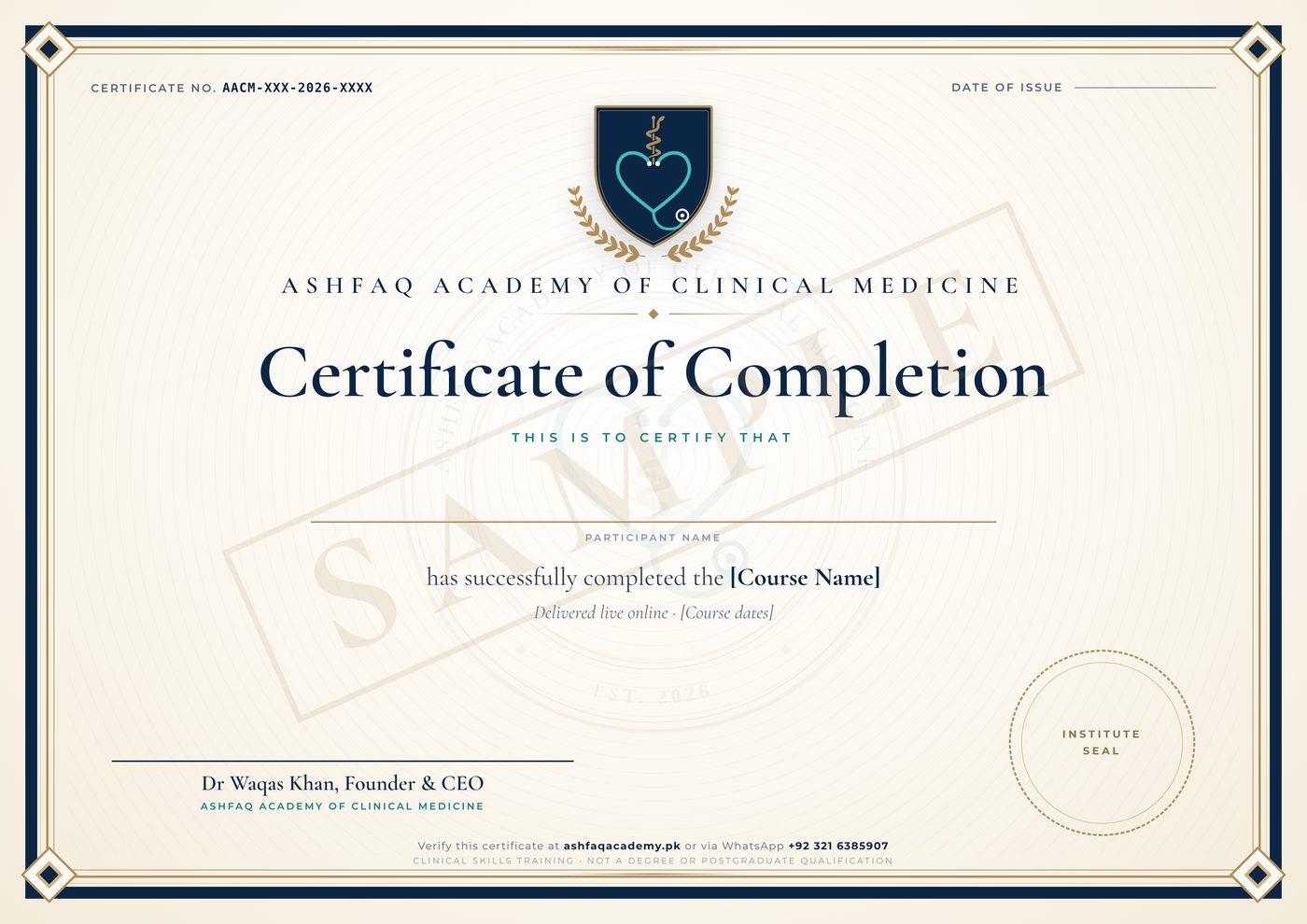
Sample certificate

Sample certificate of completion from Ashfaq Academy of Clinical Medicine, marked SAMPLE SAMPLE
Sample certificate of completion (tap to enlarge). Yours carries your name, the course dates, a certificate number, Dr Waqas Khan's signature and the institute seal. PDF
Course details

About this course

Overview

A practical masterclass on the skin and aesthetic concerns patients bring to OPD every day, from acne and pigmentation to skincare and an introduction to aesthetic procedures, taught with a strong emphasis on patient selection, safety and ethical practice.

Who it's for
  • Medical officers and GPs
  • Doctors interested in aesthetic medicine
  • Postgraduate trainees in dermatology
  • House officers
  • Nurses and allied health professionals working in skin and aesthetic clinics, within their scope of practice

Eligibility: Doctors, nurses and allied health professionals.

Dates, timings & fees
Fee: PKR 3,000. New dates will be announced soon. Register your interest and we'll message you on WhatsApp first.

Timings: 8–10 PM PKT on Zoom · usually alternate days (8 PM PKT = 7 PM UAE · 6 PM KSA · 3 PM UK (GMT) / 4 PM UK (BST)).

Dates announced soon. Every enrolled student gets 6 months of access to the session recordings. Recorded-only access is also available, with the same 6 months of access: register and choose "Recorded", and we will share the details on WhatsApp.

How you can take it
Curriculum

What we cover

Open a day or module below. Everything is still here — just collapsed so the page stays easy to scan.

  • Seven live sessions on Zoom, usually on alternate days, 8–10 PM Pakistan time — each one is one focused, continuous class, not a series of separate sessions stitched together
  • Case-based, high-yield teaching, built for OPD decision-making, not exam cramming
  • Every day moves through its conditions using the same five-lens framework, before moving to the next condition in the same category
  • Live Q&A embedded throughout every class

Day by day

  1. Day 1Foundations & Papulosquamous DisordersApproach to the dermatology patient, acne vulgaris, acne scarring, rosacea, eczema/contact/seborrhoeic dermatitis, psoriasis, and lichen planus2 hours
  2. Day 2Pigmentary, Hair & Nail DisordersFitzpatrick typing & Wood's lamp, melasma, PIH & pigmentary mimics, vitiligo, hair loss (androgenetic, telogen effluvium, alopecia areata), hirsutism, nail disorders, and acanthosis nigricans2 hours
  3. Day 3Infections, Infestations & Genital DermatosesFungal, bacterial, and viral infections, scabies & pediculosis, and common genital dermatoses2 hours
  4. Day 4Red Flags: Urticaria, Drug Reactions, Skin Cancer & Benign LesionsUrticaria & angioedema, cutaneous drug reactions (including SJS/TEN), autoimmune blistering disease, skin cancer red flags, common benign lesions patients ask about, and when to refer2 hours
  5. Day 5Aesthetic Medicine I — Injectables & Regenerative MedicineSkin ageing grading, botulinum toxin, dermal fillers, skin boosters & mesotherapy, and PRP with a full protocol2 hours
  6. Day 6Aesthetic Medicine II — Devices, Resurfacing & Scar RevisionChemical peels, microneedling, laser & light-based devices, PDO threads, and keloid/hypertrophic scar management2 hours
  7. Day 7Complex Cases, Complications, Counselling Mastery & Practice BuildingTreatment planning for combination cases, recognising and managing aesthetic complications, photoprotection counselling, counselling mastery, the ultimate FAQ vault, and building your OPD2 hours

Full curriculum

Tap a day to see every topic.

Day 1Foundations & Papulosquamous DisordersOne continuous class · 2 hours

Learning objectives

  1. Apply a structured morphological approach to any dermatology presentation
  2. Grade and manage acne vulgaris and acne scarring using an exact, escalating treatment blueprint
  3. Distinguish rosacea, eczema, and psoriasis from each other and from acne
  4. Recognise lichen planus and its mucosal and nail variants

Approach to the Dermatology Patient

Clinical Recognition
  • Core morphology vocabulary: macule, papule, plaque, nodule, vesicle, bulla, pustule, wheal — use this language for every diagnosis for the rest of the course
  • Focused history: onset, evolution, triggers, prior treatments tried, itch vs pain, systemic symptoms
  • Examination essentials: distribution pattern, symmetry, scalp/nails/mucosa — often the fastest route to diagnosis
  • When and how to use dermoscopy as a quick OPD adjunct (pigmented lesions, hair loss, scabies)

Acne Vulgaris — Comedonal to Nodulocystic

Clinical Recognition
  • Grading: comedonal (mild) → papulopustular (moderate) → nodulocystic (severe) — grade drives treatment choice
  • Distribution clues: jawline/perioral pattern suggests hormonal acne; truncal involvement signals more severe disease
Management — The Exact Blueprint by Grade
GradeFirst-Line Blueprint
ComedonalTopical retinoid at night — adapalene 0.1% (Deriva, Isotrex) or tretinoin 0.025-0.05% (A-Ret); start every other night for 2 weeks to build tolerance, then nightly
Papulopustular (mild-moderate)Topical retinoid + benzoyl peroxide 2.5-5% (Benzac) in the morning; add topical clindamycin 1% (Dalacin T) or oral doxycycline 100mg once daily for 8-12 weeks if inflammatory lesions are prominent
Nodulocystic / scarring riskOral isotretinoin (Isotane, Acnetane) 0.5-1mg/kg/day, cumulative target dose 120-150mg/kg total over the course — this is the point at which referral to, or co-management with, a dermatologist becomes appropriate given the monitoring required
Hormonal pattern in femalesCombined oral contraceptive (Diane-35, Yasmin) or spironolactone 25-100mg/day as an adjunct, particularly with jawline-predominant or premenstrual flaring acne
Red flag / safety note
  • Isotretinoin is category X in pregnancy — every female patient of reproductive age needs a documented negative pregnancy test before starting, monthly monitoring while on treatment, and counselling on two forms of contraception for the duration of treatment and one month after stopping
Patient counselling script
  • "This will take 6-8 weeks to show real improvement, and it's normal for things to look slightly worse in the first 2-3 weeks as the retinoid brings deeper comedones to the surface — that's the medicine working, not it failing."
  • "Please don't pick or pop these — I know it's tempting, but that's one of the biggest drivers of the scarring we're trying to prevent."
Frequently asked patient questions
  • "Does diet cause my acne?" — High-glycaemic-index foods and dairy have a modest, real association in some patients; worth a personal trial of reduction, but not worth an extreme restrictive diet
  • "Will it leave scars?" — Inflammatory, deeper lesions carry real scarring risk, which is exactly why earlier, adequate treatment of moderate-severe acne matters more than treating scars after the fact
  • "Will it come back after isotretinoin?" — Most patients get long-term remission after a full adequate course; a minority need a second course or ongoing topical maintenance

Acne Scarring

Clinical Recognition
  • Atrophic patterns: icepick (narrow, deep), boxcar (broad, sharp-edged), rolling (broad, sloping edges)
  • Hypertrophic/keloid scarring — more common on the trunk/jawline, raised and firm
Management & Aesthetic Intervention by Scar Type
  • Icepick scars: TCA CROSS technique (high-concentration trichloroacetic acid applied focally into the scar) is the most effective single approach; subcision has limited benefit given the narrow, deep tract
  • Boxcar scars: combination of subcision (releasing the fibrous tethering base) plus dermal filler for immediate volume, with laser resurfacing or punch excision for deeper, well-defined boxcars
  • Rolling scars: subcision is the primary technique — breaking the fibrous bands tethering the scar base to deeper tissue — often combined with microneedling or RF microneedling for overall textural improvement
  • Across 3-6 sessions; this should be stated explicitly at the first consultation, not discovered by the patient after one disappointing session
Patient counselling script
  • Scar treatment is a multi-session journey — set expectations for meaningful improvement, not erasure. Active acne must be controlled first; treating scars on actively inflamed skin worsens outcomes and risks new scarring.

Rosacea

Clinical Recognition
  • Subtypes: erythematotelangiectatic (flushing, visible vessels), papulopustular (mimics acne but no comedones), phymatous (skin thickening, classically the nose), ocular rosacea (gritty, red eyes)
  • Key differentiator from acne: absence of comedones, presence of background facial erythema and flushing
Management
  • Trigger avoidance first — spicy food, alcohol, heat, sun exposure, and topical steroids (which worsen rosacea, a genuinely common iatrogenic trap)
  • Papulopustular: topical metronidazole 0.75-1% (Rozex, Metrogyl gel) or azelaic acid 15-20% (Finacea, Azelex); topical ivermectin 1% (Soolantra) as a newer, effective alternative
  • Moderate-severe: oral doxycycline at sub-antimicrobial dose (40mg modified-release, or 100mg standard where the MR form isn't available) for its anti-inflammatory rather than antibiotic effect
  • Persistent erythema: topical brimonidine gel for same-day vasoconstriction; telangiectasia responds well to vascular laser or IPL
Frequently asked patient questions
  • "Is this just acne?" — No, and treating it as acne (especially with drying, irritating acne products) typically makes rosacea worse, not better
  • "Will it ever go away completely?" — Rosacea is a chronic, relapsing-remitting condition; the realistic goal is long-term control, not permanent cure

Eczema, Contact & Seborrhoeic Dermatitis

Clinical Recognition
  • Atopic dermatitis: flexural distribution, chronic itch-scratch cycle, personal/family atopy history
  • Contact dermatitis: sharply demarcated, linked to an identifiable exposure (allergic vs irritant)
  • Seborrhoeic dermatitis: greasy scale over the scalp, eyebrows, nasolabial folds — worsens with stress
Management
  • Atopic dermatitis: emollients as the daily cornerstone (used liberally, not just during flares); topical corticosteroid by potency matched to site and severity — mild (hydrocortisone 1%) for the face, moderate-potent (mometasone, betamethasone) for the body during flares; tacrolimus 0.03-0.1% (Protopic) or pimecrolimus (Elidel) as steroid-sparing options for the face and flexures; sedating antihistamine at night for itch-driven sleep disruption
  • Contact dermatitis: identify and eliminate the allergen/irritant — this is the actual treatment; a short course of topical steroid settles the acute reaction; patch testing for recurrent or unclear cases
  • Seborrhoeic dermatitis: ketoconazole 2% shampoo (Nizoral) used on the scalp and washed over the face/chest, or ketoconazole cream for facial involvement; a mild topical steroid for short-term use during flares; zinc pyrithione shampoo as a maintenance option
Patient counselling script
  • For atopic dermatitis: "The moisturiser isn't optional extra care — it's the actual treatment that keeps flares from happening in the first place. The steroid cream is for putting out fires; the moisturiser is for preventing them."

Psoriasis

Clinical Recognition
  • Classic plaque psoriasis: well-demarcated, silvery-scaled plaques over extensor surfaces, scalp, umbilicus; nail pitting/onycholysis
  • Other patterns: guttate (post-streptococcal, droplet lesions), inverse (flexural, minimal scale), pustular (an urgent variant)
Management
  • Limited disease: topical corticosteroid plus a vitamin D analogue, either separately or as a combination product (calcipotriol-betamethasone, e.g. Daivobet) applied once daily
  • Scalp involvement: coal tar or salicylic acid shampoo to lift scale, followed by a topical steroid solution/lotion
  • Widespread disease: narrowband UVB phototherapy where available
  • Moderate-severe or joint-involved disease: methotrexate (7.5-25mg once weekly with folic acid supplementation) is the standard systemic first-line; acitretin as an alternative; biologics are highly effective but carry significant cost and access limitations in most Pakistani practice settings, worth mentioning as an option but not a default recommendation
Patient counselling script
  • This is a chronic, genetically-influenced condition with a relapsing-remitting course — the goal is control, not cure. Ask specifically about joint pain and stiffness at every visit: undiagnosed psoriatic arthritis causes irreversible joint damage if missed, and patients rarely volunteer joint symptoms unprompted.

Lichen Planus

Clinical Recognition
  • The classic '6 Ps': Purple, Polygonal, Pruritic, Papules, Planar (flat-topped), on flexor surfaces of wrists/ankles most classically
  • Wickham's striae — fine white lines on the surface of papules, best seen with a drop of oil and a dermoscope
  • Mucosal involvement: a lacy, white, reticulate pattern on the buccal mucosa — often asymptomatic and found incidentally, but occasionally erosive and painful
  • Nail changes: longitudinal ridging, thinning, and in severe cases pterygium formation (permanent scarring fusion of the proximal nail fold to the nail bed) — a reason not to dismiss nail lichen planus as cosmetic
Management
  • Localised cutaneous disease: potent topical corticosteroid (betamethasone, clobetasol) under occlusion if needed
  • Widespread or rapidly progressive disease: a short tapering course of oral corticosteroid (prednisolone 0.5mg/kg, tapered over 4-6 weeks)
  • Oral erosive lichen planus: topical steroid in an adhesive base (triamcinolone in orabase) or a potent topical steroid mouth rinse
  • Antihistamines for itch; screen for and stop any temporally-associated new medication, since drug-induced lichenoid reactions are a genuine and easily missed mimic
Patient counselling script
  • Lichen planus is usually self-limiting, typically resolving within 1-2 years, though it can leave noticeable post-inflammatory hyperpigmentation that takes considerably longer to fade than the original lesions — set this expectation early so patients don't mistake the pigmentation for ongoing active disease.
Case-based discussion
  • A 19-year-old presents with nodulocystic acne on the jawline and chest, worse premenstrually, with 3 previous courses of topical treatment that 'didn't work.' Walk through your full assessment and treatment blueprint, including the isotretinoin conversation.
  • A 34-year-old with facial redness and visible blood vessels has been using a potent topical steroid for '3 months for her rosacea,' prescribed elsewhere, and is now worse. What happened, and how do you manage the steroid-induced component alongside the underlying rosacea?
Day 2Pigmentary, Hair & Nail DisordersOne continuous class · 2 hours

Learning objectives

  1. Apply Fitzpatrick typing to guide safe treatment selection in darker skin
  2. Distinguish melasma from post-inflammatory hyperpigmentation and its mimics
  3. Diagnose the three most common causes of hair loss from history and examination alone
  4. Recognise nail disorders and acanthosis nigricans as diagnostic windows into systemic disease

Approach to Pigmentary Disorders: Fitzpatrick Skin Typing & Wood's Lamp

Clinical Recognition
  • Fitzpatrick skin typing (I-VI) and why it matters more in our patient population than almost anywhere else — most patients fall into Type III-V, with higher post-inflammatory hyperpigmentation risk from any aggressive intervention
  • Wood's lamp as a quick OPD tool to differentiate epidermal vs dermal pigment, and to screen for fungal/pigmentary mimics

Melasma

Clinical Recognition
  • Symmetric, blotchy hyperpigmentation — centrofacial, malar, or mandibular patterns
  • Classify as epidermal, dermal, or mixed (Wood's lamp accentuation helps guide this)
  • Identify drivers: sun exposure, hormonal triggers (pregnancy, OCPs), family history
Management — The Exact Blueprint
  • First-line topical: triple combination cream (hydroquinone 4% + tretinoin 0.05% + a mild steroid, e.g. Tri-Luma) applied at night for up to 8 weeks (its licensed course), then step down to hydroquinone alone or a non-hydroquinone maintenance agent
  • Non-hydroquinone options for maintenance or hydroquinone-intolerant patients: azelaic acid 20%, tranexamic acid (topical or oral 250mg BD in resistant cases, screened for contraindications), kojic acid, and niacinamide-based combinations
  • Adjunct in-office procedures: superficial chemical peels (glycolic, mandelic) or low-fluence Q-switched Nd:YAG toning — always as an ADJUNCT to topical therapy and strict photoprotection, never as a replacement for either
  • Non-negotiable throughout treatment: daily broad-spectrum sunscreen, reapplied — melasma treated without genuine sun discipline will not hold its improvement
Patient counselling script
  • "Melasma is a condition we control, not cure — the goal is getting it as light as possible and keeping it there, and that second part depends entirely on your sunscreen habit, not on which cream we use."
Frequently asked patient questions
  • "Will it go away after pregnancy/stopping the pill?" — Often improves, but doesn't always fully resolve, and any UV exposure afterward can reactivate it
  • "Can I get a peel or laser for this?" — Yes, but only as an adjunct to topical treatment and photoprotection, and the wrong laser parameters in melasma can genuinely worsen it — this is not a condition for aggressive, high-energy treatment

Post-Inflammatory Hyperpigmentation & Pigmentary Mimics

Clinical Recognition
  • PIH follows a clear inflammatory trigger (acne, eczema, trauma) — distribution matches the prior lesion
  • Differentiate from melasma (symmetric, no preceding inflammation) and tinea versicolor (fine scale, Wood's lamp fluorescence, KOH positive)
Management
  • Treat the underlying inflammatory condition first — PIH will not resolve while the trigger remains active
  • Topical: azelaic acid, niacinamide, or a short course of hydroquinone for stubborn cases; retinoids accelerate epidermal turnover and fading
  • Strict sun protection — PIH darkens rapidly with UV exposure and this is the single most common reason it fails to fade

Vitiligo

Clinical Recognition
  • Well-demarcated, depigmented (not hypopigmented) macules/patches; classic sites — face, hands, genitalia, periorificial
  • Signs of activity: Koebner phenomenon, confetti-like lesions, trichrome border — important for treatment planning and counselling
Management
  • Localised, stable disease: potent topical corticosteroid or topical tacrolimus 0.1% (Protopic), particularly for the face where steroid atrophy risk is higher
  • Widespread or active disease: narrowband UVB phototherapy is the standard first-line systemic option where available; oral mini-pulse corticosteroids can arrest rapidly progressive disease
  • Camouflage and depigmentation (for extensive, refractory facial disease) are legitimate options to discuss once medical treatment has been given a fair trial
Patient counselling script
  • This is a genuine autoimmune condition, not something the patient did or ate — a surprising number of patients arrive having been told otherwise by well-meaning family. Treatment response is slow (months, not weeks) and repigmentation, when it happens, starts around hair follicles as small dots before merging — showing a patient this pattern early can keep them engaged with a slow-responding treatment.

Hair Loss — Androgenetic Alopecia, Telogen Effluvium, Alopecia Areata

Clinical Recognition
  • Androgenetic alopecia: gradual, patterned thinning (frontotemporal/vertex in men, diffuse crown thinning with a preserved frontal line in women)
  • Telogen effluvium: diffuse shedding 2-3 months after a triggering event (illness, surgery, postpartum, crash dieting, severe stress)
  • Alopecia areata: well-defined, smooth round patches of hair loss, often sudden onset; exclamation-mark hairs at the margin
  • Basic work-up to consider: ferritin, thyroid profile, and a directed history for triggers/nutritional status
Management — The Exact Blueprint
DiagnosisFirst-Line Treatment
Androgenetic alopeciaTopical minoxidil 5% solution/foam once or twice daily (both sexes); oral finasteride 1mg/day in men (screen for and counsel on sexual side effects); spironolactone as an off-label option in women with a hormonal pattern
Telogen effluviumIdentify and correct the trigger; iron and vitamin D repletion if deficient; reassurance that this self-resolves over 6-12 months once the trigger is addressed — minoxidil can shorten the course but is not strictly necessary
Alopecia areata (limited patches)Intralesional triamcinolone acetonide (5-10mg/mL, injected at multiple points within the patch) is first-line; topical potent steroids as an alternative for needle-averse patients or young children
Alopecia areata (extensive/rapidly progressive)Referral for consideration of contact immunotherapy, systemic steroids, or JAK inhibitor therapy where accessible
Frequently asked patient questions
  • "Is this genetic and permanent?" (androgenetic alopecia) — Yes, it's progressive without treatment, but both minoxidil and finasteride genuinely slow and partially reverse it if started before extensive miniaturisation has occurred — earlier treatment gives meaningfully better results
  • "Will my hair grow back?" (telogen effluvium) — Almost always yes, once the trigger resolves, though it can take several months to become visually obvious

Hirsutism — Brief Approach

Clinical Recognition
  • Male-pattern terminal hair growth in women — screen for PCOS and other endocrine causes when associated with irregular cycles, acne, or weight changes
Management
  • Address the underlying cause where identified (PCOS management, combined oral contraceptive, spironolactone 50-200mg/day for the androgen-driven component)
  • Cosmetic hair removal (laser hair reduction being the most durable option) for direct symptomatic management alongside medical treatment

Nail Disorders

Clinical Recognition
  • Onychomycosis: thickened, discoloured (yellow-brown), crumbly nail plate, usually starting distally; confirm with KOH/culture before committing to months of oral treatment — this is genuinely worth doing, since several look-alike conditions don't need antifungals at all
  • Psoriatic nail disease: pitting, onycholysis, oil-drop discolouration, subungual hyperkeratosis — a marker worth specifically linking back to joint-disease screening
  • Paronychia: acute (painful, red, swollen, often with pus — usually bacterial) vs chronic (repeated wet work, candida involvement, affects the proximal nail fold over weeks)
  • Ingrown toenail (onychocryptosis): lateral nail edge penetrating the surrounding skin, usually the great toe, causing pain and secondary infection
Management
  • Onychomycosis: oral terbinafine 250mg/day for 6 weeks (fingernails) or 12 weeks (toenails) is first-line given superior cure rates over topical treatment alone; topical amorolfine or ciclopirox lacquer for mild distal disease or where oral treatment is contraindicated; check LFTs before and during oral terbinafine courses beyond a few weeks
  • Chronic paronychia: keep hands dry, avoid prolonged wet work, topical antifungal/steroid combination since candida and low-grade eczema often coexist here
  • Acute paronychia: warm soaks, incision and drainage if a clear abscess has formed, oral antibiotics (cloxacillin or cefalexin) if cellulitis is present
  • Ingrown toenail: conservative measures (proper trimming technique, cotton wisp elevation) for mild cases; partial nail avulsion with phenolisation of the nail matrix for recurrent cases

Acanthosis Nigricans

Clinical Recognition
  • Velvety, hyperpigmented, thickened plaques — classically the neck, axillae, and other flexural areas
  • The single most important recognition point: this is a cutaneous marker of insulin resistance, not a primary skin disease — the skin finding should trigger a metabolic work-up, not just a request for a cosmetic fix
  • Rapid onset, widespread distribution, or involvement of unusual sites (mucosa, palms) in an adult raises concern for an underlying malignancy (malignant acanthosis nigricans) and warrants prompt further investigation
Management
  • Screen for and address the underlying driver: fasting glucose/HbA1c, and in adolescent girls, a PCOS work-up if the clinical picture fits — this is genuinely the treatment, not a cosmetic cream
  • Weight management where relevant meaningfully improves the skin findings over time
  • Topical retinoids or alpha-hydroxy acids can offer modest cosmetic improvement in texture and pigmentation once the underlying driver is being addressed, but should never be offered as a substitute for the metabolic work-up
Clinical pearls & practical tips
  • A teenager brought in by a parent purely for 'the dark neck' is one of the highest-value opportunistic screening moments in general OPD — this single visual finding, followed through properly, catches insulin resistance years before it would otherwise present.
Case-based discussion
  • A 28-year-old woman presents with symmetric brown patches across both cheeks that started during her second pregnancy 2 years ago and haven't faded. Walk through your classification, treatment blueprint, and the sunscreen conversation.
  • A 16-year-old girl is brought in by her mother 'just for the dark neck' — she has no other complaints. What do you look for, what do you ask, and what do you tell the family?
Day 3Infections, Infestations & Genital DermatosesOne continuous class · 2 hours

Learning objectives

  1. Diagnose and treat fungal, bacterial, and viral skin infections with exact regimens and durations
  2. Recognise scabies and pediculosis, including household contact management
  3. Approach common genital dermatoses in general OPD without unnecessary embarrassment for the patient or the clinician

Fungal Infections — Tinea & Candidiasis

Clinical Recognition
  • Annular, scaly, advancing-edge plaques with central clearing — classic tinea morphology
  • Tinea capitis: scaling with or without patchy alopecia in children — always consider when a child presents with patchy hair loss
  • Candidiasis: beefy-red, macerated plaques with satellite pustules in moist, intertriginous areas
  • KOH microscopy as a quick, high-yield OPD confirmatory test
Management — The Exact Blueprint
  • Tinea corporis/cruris (localised): topical terbinafine 1% or clotrimazole 1%, twice daily for 2-4 weeks — continue for at least a week past visible clearance to prevent early relapse, a genuinely common cause of treatment 'failure'
  • Tinea pedis: as above, plus keeping feet dry and treating any concurrent onychomycosis, which otherwise reseeds the skin infection
  • Widespread, recurrent, or steroid-modified tinea (tinea incognito, from prior inappropriate steroid use): oral terbinafine 250mg/day for 2-4 weeks, or oral itraconazole as an alternative — note that terbinafine-resistant dermatophytosis (Trichophyton indotineae) is now widespread in South Asia, so itraconazole is often preferred in Pakistan; ask about adherence and prior treatment, and consider mycology if a course fails
  • Tinea capitis: ALWAYS requires an oral antifungal — topical treatment alone cannot penetrate the hair follicle; griseofulvin 20mg/kg/day for 6-8 weeks remains a standard, well-tolerated choice in children, with oral terbinafine as a shorter-course alternative
  • Candidiasis: topical clotrimazole or nystatin cream for skin; keep the area dry; address contributing factors (diabetes, obesity, incontinence, recent antibiotic use)
Clinical pearls & practical tips
  • Tinea that looks unusual, less scaly, or oddly well-controlled-but-not-clearing has often already been treated with a topical steroid-antifungal combination cream (widely and inappropriately sold over the counter) — always ask specifically about prior self-treatment before assuming your diagnosis or your antifungal choice is wrong.

Bacterial Infections — Impetigo, Folliculitis, Cellulitis, Abscess

Clinical Recognition
  • Impetigo: honey-coloured crusted erosions, classically perioral/periorificial in children
  • Folliculitis: follicular pustules; cellulitis: spreading erythema, warmth, tenderness, with or without fever; abscess: fluctuant, tender swelling
Management
  • Impetigo (localised): topical mupirocin 2% or fusidic acid, three times daily for 5-7 days
  • Impetigo (widespread) or folliculitis not responding to topical treatment: oral cephalexin (25-50mg/kg/day) or cloxacillin (50-100mg/kg/day in 4 divided doses) in children, for 7 days
  • Cellulitis (uncomplicated, no systemic toxicity): oral cloxacillin or cephalexin for 5-7 days, with a clear review plan if not improving within 48-72 hours
  • Abscess: incision and drainage is the primary treatment — antibiotics alone rarely resolve a formed abscess without drainage
Red flag / safety note
  • Rapidly spreading cellulitis, systemic toxicity (fever, hypotension, confusion), facial/periorbital cellulitis, or any suspicion of necrotising infection — these need urgent referral/admission, not OPD management

Viral Infections — Warts, Molluscum, HSV, Herpes Zoster

Clinical Recognition
  • Verrucae: rough, hyperkeratotic papules, often with thrombosed capillary puncta
  • Molluscum contagiosum: dome-shaped, umbilicated papules — common in children and immunosuppressed adults
  • Herpes simplex: grouped vesicles on an erythematous base, recurrent at the same site
  • Herpes zoster: dermatomal, unilateral vesicular eruption — recognise early for antiviral timing
Management
  • Verrucae: topical salicylic acid preparations as first-line for home use; cryotherapy in-office for resistant lesions, repeated every 2-3 weeks
  • Molluscum: often self-limiting over months and can genuinely be watched in an asymptomatic, cooperative child; curettage or cryotherapy for persistent, spreading, or cosmetically bothersome lesions
  • Herpes simplex: oral aciclovir 200mg five times daily or 400mg three times daily (or valaciclovir 500mg BD) for 5 days, started as early as possible in the prodrome/first 72 hours for maximum benefit; suppressive daily therapy for patients with frequent recurrences
  • Herpes zoster: oral aciclovir 800mg five times daily (or valaciclovir 1g TDS) for 7 days, ideally started within 72 hours of rash onset; adequate analgesia given the genuine risk of post-herpetic neuralgia
Red flag / safety note
  • Herpes zoster involving the eye/nasal tip (ophthalmic zoster, suggested by a vesicle at the tip of the nose — Hutchinson's sign) needs urgent ophthalmology referral given the risk of vision loss

Scabies & Pediculosis

Clinical Recognition
  • Scabies: intensely pruritic, worse at night; burrows in finger webs, wrists, genitalia; look for household contacts with similar symptoms
  • Pediculosis: visible nits/lice on the scalp or body hair, associated itching
Management
  • Scabies: permethrin 5% cream applied neck-to-toe (whole body in infants), left on for 8-12 hours, repeated after 1 week to catch newly-hatched mites; oral ivermectin (200mcg/kg, repeated after 1-2 weeks) as an alternative for extensive disease, crusted scabies, or outbreak settings
  • ALL household and close contacts must be treated SIMULTANEOUSLY, symptomatic or not — this is the single most common reason for treatment 'failure,' which is usually reinfestation, not resistance
  • Wash all bedding and clothing used in the preceding 3 days in hot water, or seal in a plastic bag for 72 hours if washing isn't practical
  • Pediculosis: permethrin 1% shampoo/lotion, or malathion as an alternative; wet-combing with a fine-toothed comb as an adjunct; repeat treatment at day 7-9 to catch newly hatched lice
Patient counselling script
  • "The itching can genuinely continue for 2-3 weeks after successful treatment — this is a normal allergic reaction to the dead mites, not a sign the treatment failed. Don't repeat treatment early just because the itch hasn't stopped, unless new burrows are appearing."

Common Genital Dermatoses

Clinical Recognition
  • Lichen sclerosus: well-demarcated, white, atrophic ('cigarette-paper') plaques, classically in a figure-of-eight pattern around the vulva and perianal area in women, or affecting the glans/foreskin (balanitis xerotica obliterans) in men — itching, soreness, and in men, progressive phimosis are the usual presenting complaints
  • Balanitis/balanoposthitis: redness, soreness, and discharge of the glans (with the foreskin in balanoposthitis) — candidal (satellite lesions, common in uncircumcised men and diabetics), irritant/contact, or bacterial causes
  • Vulvovaginal candidiasis: itching, soreness, thick white discharge — extremely common and frequently self-diagnosed correctly by patients, though recurrent cases deserve a genuine work-up
  • Genital psoriasis: well-demarcated, often less scaly than elsewhere given the moist environment — easily misdiagnosed as a fungal infection if the possibility isn't actively considered
Management
  • Lichen sclerosus: ultra-potent topical corticosteroid (clobetasol propionate) is first-line and highly effective if used correctly and for long enough — this is not a condition to under-treat given the genuine risk of scarring and, in longstanding vulvar disease, a small but real increased risk of squamous cell carcinoma, which is why follow-up matters beyond just symptom relief
  • Candidal balanitis: topical clotrimazole; treat and screen for diabetes if recurrent, since recurrent candidal balanitis can be the first presenting sign of undiagnosed diabetes in a man with no other symptoms
  • Genital psoriasis: a mild-moderate topical steroid, used for shorter courses given the increased absorption in genital skin; avoid the more irritating vitamin D analogues in this location
Clinical pearls & practical tips
  • A brief, matter-of-fact, purely clinical approach to genital examination — explaining what you're going to look at and why before you do it — does more to put a patient at ease than any amount of apologising or hesitation. Patients take their cue from the clinician's comfort level.
Case-based discussion
  • A 7-year-old boy has a well-defined, scaly patch on his scalp with broken-off hair stubs, unresponsive to an antifungal cream from a pharmacy 2 weeks ago. What's actually going on, and what's your management blueprint?
  • A 45-year-old man presents with recurrent balanitis for the third time this year despite topical clotrimazole each time. What are you specifically screening for, and why?
Day 4Red Flags: Urticaria, Drug Reactions, Skin Cancer & Benign LesionsOne continuous class · 2 hours

Learning objectives

  1. Manage acute and chronic urticaria, and recognise angioedema requiring emergency care
  2. Distinguish a benign drug rash from an SJS/TEN-spectrum emergency
  3. Apply the ABCDE rule and recognise BCC, SCC, and actinic keratosis with confidence
  4. Correctly identify the common benign lesions patients ask about, without over- or under-investigating them
  5. Recognise autoimmune blistering disease and build a personal referral threshold across all of dermatology OPD

Urticaria & Angioedema

Clinical Recognition
  • Wheals: itchy, raised, evanescent (individual lesions resolve within 24 hours) — the key differentiator from other rashes
  • Acute (under 6 weeks) vs chronic spontaneous urticaria (6 weeks or more)
  • Angioedema: deeper swelling, often lips/eyelids/tongue — assess airway involvement urgently
Management — The Exact Blueprint
  • Acute urticaria: a second-generation, non-sedating antihistamine (cetirizine 10mg, loratadine 10mg, or fexofenadine 180mg) once daily; identify and remove any obvious trigger
  • Refractory acute symptoms: updose the same antihistamine up to 4× the standard dose before switching agents — this is standard, evidence-based practice and genuinely under-used in general OPD, where clinicians often switch antihistamines rather than increasing the dose of one that's partially working
  • Chronic spontaneous urticaria: updosed second-generation antihistamine as above, given consistently rather than as-needed; refer for consideration of omalizumab if there's no adequate response after 2-4 weeks of updosed antihistamine — a highly effective option where accessible, though cost is a genuine limiting factor in most Pakistani practice settings
  • Avoid long-term systemic corticosteroids for chronic urticaria — a short course can settle a severe acute flare, but repeated or prolonged steroid courses are not appropriate maintenance treatment
  • Isolated recurrent angioedema without wheals: consider ACE-inhibitor-induced angioedema (stop the drug) and, if truly recurrent and unexplained, hereditary angioedema (C1 esterase inhibitor deficiency) as a differential worth referring for
Red flag / safety note
  • Angioedema with tongue/throat swelling, stridor, or breathing difficulty is anaphylaxis until proven otherwise — this is an emergency requiring immediate epinephrine and emergency care, not an OPD visit

Cutaneous Drug Reactions

Clinical Recognition
  • Morbilliform/exanthematous drug rash: the most common pattern, widespread maculopapular eruption, usually benign
  • Severe cutaneous adverse reactions (SJS/TEN spectrum): mucosal involvement, skin pain, blistering, a positive Nikolsky sign — recognise this distinction immediately
Management
  • Morbilliform rash: if the causative drug is not essential, stop it; symptomatic relief with oral antihistamines and, if needed, a short course of a mild-moderate topical steroid; the rash typically fades over 1-2 weeks even without any specific treatment
  • SJS/TEN spectrum: STOP the suspected drug immediately — this single action is the most important determinant of outcome and should not wait for a specialist opinion; admit for supportive, burns-unit-style care (fluid/electrolyte management, wound care, infection surveillance); SCORTEN criteria are used by the admitting team to estimate prognosis
Red flag / safety note
  • Any mucosal involvement, skin pain out of proportion to appearance, blistering/sloughing, or systemic symptoms with a recent new drug — treat as an SJS/TEN-spectrum emergency: stop the culprit drug and refer/admit immediately. Do not manage this in routine OPD.
Patient counselling script
  • Document and clearly communicate the culprit drug to the patient (and any other treating physicians, in writing where possible) to prevent re-exposure — this matters for every drug reaction, not only the severe ones, since even a morbilliform rash predicts a similar or worse reaction on re-challenge.

Skin Cancer Recognition — The Red Flag Most OPD Curricula Skip

Clinical Recognition — Melanoma
  • The ABCDE rule for any pigmented lesion: Asymmetry, Border irregularity, Colour variation (multiple shades within one lesion), Diameter over 6mm, Evolving (any change in size, shape, colour, or symptoms — arguably the single most important letter, since it applies regardless of the lesion's size or appearance at any one visit)
  • The 'ugly duckling sign': a pigmented lesion that looks different from the patient's other moles is a more reliable real-world screening tool than trying to apply all five ABCDE criteria to every single lesion in a patient with many naevi
  • New pigmented lesions after age 40, or any lesion under a nail (subungual, especially with pigment extending onto the surrounding skin — Hutchinson's sign), deserve particular attention
Clinical Recognition — Basal Cell Carcinoma (BCC)
  • The most common skin cancer overall; slow-growing, on sun-exposed skin (face is classic)
  • Classic appearance: a pearly or translucent papule/nodule with a rolled border and visible telangiectasia over the surface; central ulceration in longstanding lesions gives the traditional 'rodent ulcer' description
  • Rarely metastasises, but left untreated causes progressive local tissue destruction — this is a 'don't need to panic, but do need to act' lesion, not an emergency and not something to ignore
Clinical Recognition — Squamous Cell Carcinoma (SCC) & Actinic Keratosis
  • Actinic keratosis: the precursor lesion — rough, scaly, erythematous patches on chronically sun-exposed skin, often easier to feel (a sandpaper texture) than to see clearly
  • SCC: a scaly, crusted, or ulcerated papule/plaque, growing faster than BCC, with genuinely higher metastatic potential — particularly on the lip and ear, where a lower threshold for biopsy and referral is warranted
Management
  • Any lesion meeting ABCDE criteria, showing the ugly duckling sign, or otherwise causing genuine clinical suspicion for melanoma: refer for an EXCISIONAL biopsy with a narrow margin — never a shave biopsy, which can compromise accurate staging (Breslow thickness) if the lesion proves malignant
  • Suspected BCC or SCC: biopsy (punch or shave is acceptable here, unlike for suspected melanoma) to confirm before definitive treatment, which is typically surgical excision; Mohs micrographic surgery for high-risk or cosmetically sensitive sites where available
  • Actinic keratosis: cryotherapy for isolated lesions; topical 5-fluorouracil or diclofenac gel for field treatment of multiple lesions across a sun-damaged area
Red flag / safety note
  • Any lesion that is changing, bleeding, non-healing beyond 4 weeks, or meets ABCDE criteria needs biopsy or urgent dermatology referral — this is the single highest-stakes 'don't miss' category in an entire dermatology OPD career, and the cost of a missed melanoma is measured in years of life, not just a delayed diagnosis.
Clinical pearls & practical tips
  • Get into the habit of a genuine full-skin check — not just the lesion the patient points to — in any patient over 40, particularly with a history of significant sun exposure. Patients very often present about one cosmetic concern while sitting on a genuinely suspicious lesion they haven't mentioned because it doesn't bother them.

Common Benign Lesions Patients Ask About

Clinical Recognition
LesionRecognitionKey Point
Seborrhoeic keratosis'Stuck-on,' waxy or warty, well-demarcated, tan to black; increases with ageCan be heavily pigmented and mimic melanoma — if there's genuine doubt, biopsy rather than assume; otherwise reassure and remove only if symptomatic or cosmetically bothersome
DermatofibromaFirm, often pigmented papule, usually on the legs; a history of minor trauma or an insect bite is commonThe 'dimple sign' — the lesion dimples inward on lateral pinching — is a genuinely reliable bedside confirmatory test
Skin tags (acrochordons)Soft, pedunculated, skin-coloured to brown, in flexures and on the neckSimple snip or cautery removal if bothersome; multiple tags in a younger or overweight patient is worth a passing mention of insulin resistance risk
Cherry angiomaSmall, smooth, bright red-to-purple papules; increase in number with ageEntirely benign vascular proliferation; laser or electrocautery for cosmetic removal if requested
MiliaTiny (1-2mm), firm, white, keratin-filled cysts, classically around the eyesSimple needle extraction; common and harmless, but patients often assume they're 'whiteheads' and worth explaining the difference
Sebaceous hyperplasiaYellowish, soft, umbilicated papules on the face in older adultsThe umbilicated centre and yellowish colour help distinguish this from BCC, though genuine diagnostic uncertainty warrants biopsy rather than guessing
Frequently asked patient questions
  • "Is this dangerous?" — For every lesion on this list, the honest answer is no, provided the diagnosis is confidently benign on examination; the skill this section builds is knowing when you're confident enough to say that outright, and when a lesion's appearance genuinely warrants a biopsy instead of reassurance

Autoimmune Blistering Disease

Clinical Recognition
  • Pemphigus vulgaris: FLACCID bullae that rupture easily, leaving painful erosions; a POSITIVE Nikolsky sign (lateral pressure on normal-appearing skin causes it to shear off); oral mucosal erosions are very often the FIRST presenting sign, sometimes preceding skin lesions by weeks to months — a patient with persistent, unexplained oral erosions deserves this on the differential, not just aphthous ulcers or a viral cause
  • Bullous pemphigoid: TENSE bullae (a genuinely useful bedside contrast to pemphigus's flaccid ones), typically in an OLDER patient population, NEGATIVE Nikolsky sign, and comparatively LESS mucosal involvement — generally a less severe disease course than pemphigus vulgaris
Management
  • Both conditions require biopsy (including direct immunofluorescence) for definitive diagnosis — this is specialist referral territory, but recognising the pattern in OPD and referring promptly, rather than treating empirically as an infection or a drug reaction, is what changes the outcome
  • Systemic corticosteroids are the mainstay of initial treatment for both, often alongside a steroid-sparing immunosuppressant for longer-term control, managed jointly with dermatology
Clinical pearls & practical tips
  • The flaccid-vs-tense bullae distinction, plus the Nikolsky sign, is genuinely one of the highest-yield two-second bedside tests in all of dermatology — it takes moments to check and meaningfully narrows a differential that otherwise looks similar at first glance.

When to Refer / Safety-Netting in Dermatology OPD

Management
  • Build a personal referral threshold for: suspected SJS/TEN, necrotising soft tissue infection, ophthalmic zoster, any lesion meeting ABCDE criteria or otherwise atypical/changing, suspected autoimmune blistering disease, and any systemically unwell patient
  • Safety-netting language for the patient: clear instructions on what symptoms should bring them back immediately, not at the next routine visit
Case-based discussion
  • A 58-year-old farmer presents with a slowly growing, pearly nodule on his nose with visible tiny blood vessels over its surface, present for 'maybe a year.' Walk through your recognition, biopsy decision, and referral pathway.
  • A 30-year-old presents with persistent, painful oral erosions for 6 weeks, treated twice already as 'oral thrush' and once as herpes, with no improvement. What are you now considering, and what's your next step?
Day 5Aesthetic Medicine I — Injectables & Regenerative MedicineOne continuous class · 2 hours

Learning objectives

  1. Grade skin ageing and match it to the right intervention conversation
  2. Select botulinum toxin dosing and injection points by facial zone, and recognise complications early
  3. Choose the right filler type, technique, and zone — and manage vascular occlusion as the genuine emergency it is
  4. Understand skin boosters and mesotherapy as a distinct category from volumising filler
  5. Run a complete, evidence-based PRP protocol for both facial and hair indications

Skin Ageing — Recognition & Grading

Clinical Recognition
  • Intrinsic ageing (chronological, genetic) vs extrinsic ageing (photoageing, smoking, pollution) — the distinction shapes counselling and treatment choice
  • Glogau photoageing scale as a quick, practical grading framework to anchor intervention discussions: Type I (mild — no wrinkles, early photoageing), Type II (moderate — wrinkles in motion, early lentigines), Type III (advanced — wrinkles at rest, visible telangiectasia, dyschromia), Type IV (severe — wrinkles throughout, no normal skin colour, prior skin cancers)
  • Matching intervention intensity to grade prevents both under-treating a genuinely advanced case with topicals alone, and over-treating a mild case with unnecessarily aggressive procedures

Botulinum Toxin

Clinical Recognition
  • Indications: dynamic wrinkles (glabellar frown lines, horizontal forehead lines, crow's feet), select therapeutic uses (axillary/palmar hyperhidrosis, masseter hypertrophy for facial slimming and bruxism) — know the indication before the technique
  • Products available in Pakistan: onabotulinumtoxinA (Botox), abobotulinumtoxinA (Dysport), incobotulinumtoxinA (Xeomin), and several regional brands (Neuronox, Botulift) at a lower price point — dosing UNITS are NOT interchangeable between brands, and this is a genuinely common source of dosing error when switching products
Management — Exact Dosing Blueprint by Zone (onabotulinumtoxinA units)
ZoneTypical DoseKey Technique Point
Glabella (frown lines)20-25 units across 5 injection pointsStay at least 1cm above the bony orbital rim to avoid diffusion into levator palpebrae and resulting ptosis
Forehead (horizontal lines)10-20 units, dosed conservativelyUndertreat on the first visit if unsure — a heavy, 'frozen' brow is far easier to avoid than to fix, and brow ptosis is the classic overtreatment complication here
Crow's feet12-15 units per side, 3 pointsInject superficially and stay lateral to the orbital rim to avoid affecting the zygomaticus and causing an asymmetric smile
Masseter (slimming/bruxism)20-30 units per side, 2-3 pointsConfirm the injection is within muscle belly by having the patient clench; effect on facial width takes 4-8 weeks to become visible, and this should be stated upfront
Axillary hyperhidrosis50 units per side, gridded 1-1.5cm apartConfirm the treatment area first with a starch-iodine test where available; effect is genuinely dramatic and long-lasting (6-12 months) compared with facial dosing
  • Onset of visible effect: 3-7 days, with full effect at 2 weeks — always schedule the review appointment at 2 weeks, not before, since touch-ups assessed too early lead to unnecessary re-treatment
  • Duration: typically 3-4 months for facial dosing, longer for hyperhidrosis
Red flag / safety note
  • Ptosis (brow or eyelid) is the complication to actively prevent through correct injection depth and distance from the orbital rim, not just manage after the fact — once it occurs, it resolves only with time (usually 4-6 weeks) as the toxin wears off; apraclonidine eye drops can provide temporary symptomatic improvement of eyelid ptosis (not brow ptosis) by stimulating Müller's muscle, but there is no way to reverse the toxin itself
Patient counselling script
  • "This isn't permanent — expect it to soften gradually over 3-4 months, and most patients settle into a rhythm of treatment two to three times a year." Advise against lying flat, rubbing the treated area, or vigorous exercise for the rest of the treatment day, to reduce the theoretical risk of diffusion.
Frequently asked patient questions
  • "Will I look frozen?" — Not with correctly dosed, conservative treatment; overtreatment (and often self-inflicted by requesting 'as much as possible') is what causes the frozen look, not the treatment itself
  • "Is it safe to do this regularly for years?" — Yes; there's no evidence of cumulative harm from repeated, correctly-spaced treatment over many years

Dermal Fillers

Clinical Recognition
  • Categories: hyaluronic acid (most common, reversible) vs other biostimulatory or semi-permanent agents — reversibility is a key patient-safety and counselling point
  • Indications by facial zone: volume loss (cheeks, temples), lip enhancement, nasolabial folds, marionette lines, jawline contouring
Management — Product Selection by Zone
  • Hyaluronic acid (HA): selected by viscosity/G-prime for the zone — a lower-viscosity, softer HA for lips and the tear trough; a higher-viscosity, more structural HA for cheek and jawline volumising. The genuine safety advantage of HA fillers is reversibility with hyaluronidase, which is exactly why they remain first-line for most indications in a general aesthetic practice.
  • Calcium hydroxylapatite (CaHA): provides both immediate volume and a genuine biostimulatory collagen effect over subsequent months — well suited to the jawline and hands, but NOT reversible with hyaluronidase, which matters directly for complication management.
  • Poly-L-lactic acid (PLLA): purely biostimulatory with no immediate volumising effect — results build gradually across 2-3 sessions over months as the body lays down new collagen, and this timeline needs to be set clearly at consultation so a patient doesn't judge the outcome after session one.
  • Permanent fillers: not recommended — the complication profile (chronic granuloma formation, migration, and the simple fact that a complication cannot be reversed years later) outweighs the appeal of a 'one-time' treatment.
  • Cannula technique is preferred over a sharp needle in higher-risk vascular zones (tear trough, temple, nose) given its comparatively lower vascular injury risk, though it does not eliminate that risk entirely
Red flag / safety note
  • Vascular occlusion is the critical complication to recognise immediately: sudden severe pain, skin blanching or a mottled/livedo pattern, or delayed bruising disproportionate to the procedure. This is a TIME-SENSITIVE emergency. Immediate management: STOP injecting, apply warm compresses, massage the area, and inject hyaluronidase (Hyalase) generously and immediately into and around the affected area if an HA filler was used — do not wait to 'see if it improves.' Non-HA fillers have no equivalent antidote, which is exactly why product choice in high-risk zones deserves real thought upfront.
  • Vision loss from retrograde embolisation is rare but catastrophic, most associated with glabellar and nasal injections — know this risk exists specifically in these zones and inject with correspondingly greater caution (slow injection, low pressure, aspiration where feasible, cannula preferred).
  • Other complications to recognise early: infection, delayed-onset nodule/granuloma formation (weeks to months later — don't dismiss a new lump simply because 'it's been too long since the filler'), and allergic reaction.
Patient counselling script
  • Every filler consultation needs explicit informed consent covering realistic outcome, expected duration (typically 9-18 months for most HA fillers, zone-dependent), and an honest account of the vascular occlusion risk — this is not a conversation to abbreviate because it might make the patient nervous; an informed, calm patient is exactly what you want if a complication does occur.
Frequently asked patient questions
  • "Can this be dissolved if I don't like it?" — Yes, for HA fillers, using hyaluronidase; this is worth mentioning proactively, since it meaningfully reduces a hesitant patient's anxiety about committing to treatment
  • "Will people be able to tell I've had filler?" — Correctly dosed, zone-appropriate filler aims for the patient looking like a well-rested version of themselves, not visibly 'done' — this is a legitimate expectation to set explicitly, not just assume the patient shares

Skin Boosters & Mesotherapy

Clinical Recognition
  • A genuinely distinct category from volumising filler — the goal here is skin QUALITY (hydration, elasticity, fine textural improvement), not shape or volume change, and this distinction is the single most important thing to get across to a patient who may arrive expecting filler-like results
  • Skin boosters (Profhilo, Restylane Vital/Skinbooster range) use non-cross-linked or minimally cross-linked hyaluronic acid, injected superficially in a grid of small bolus points or micro-droplets rather than the linear threading or deeper bolus technique used for volumising filler
  • Mesotherapy uses a broader cocktail — typically combinations of hyaluronic acid, vitamins, amino acids, and antioxidants — delivered via multiple superficial micro-injections across the treatment area
Management
  • Standard course: 2-3 sessions spaced 3-4 weeks apart for the initial series, with maintenance sessions every 6-9 months thereafter
  • Common treatment areas: face, neck, décolletage, and backs of the hands — areas where skin quality/laxity is the primary concern rather than volume loss
  • Can be combined with volumising filler in the same treatment plan — skin boosters address surface quality while structural filler addresses volume, and these are complementary, not competing, treatments
Patient counselling script
  • "This isn't going to change your shape or add volume — think of it as a deep, sustained hydration treatment for the skin itself. You'll likely notice smoother texture and a healthier glow before you notice any dramatic 'lifted' appearance."

Platelet-Rich Plasma (PRP)

Clinical Recognition
  • Autologous, growth-factor-rich plasma prepared from the patient's own blood — genuinely low allergic/rejection risk given the autologous source
  • Two primary aesthetic indications: facial skin rejuvenation (often combined with microneedling for delivery) and androgenetic alopecia (as an adjunct to topical/oral medical therapy, not a replacement for it)
Management — The Full Protocol
  • Preparation: draw 10-20mL of venous blood into citrate tubes; centrifuge (a single-spin protocol is standard for most in-office systems; some protocols use a double spin for a more concentrated platelet yield) to separate and concentrate the platelet-rich plasma fraction from red cells and platelet-poor plasma
  • Facial delivery: apply via microneedling (typically 0.5-1.5mm depth depending on the zone and indication) immediately after PRP application, allowing the growth factors direct access to the dermis; alternatively, fine-needle micro-injection across the treatment area
  • Facial protocol: 3-4 sessions at 4-week intervals for the initial course, with a maintenance session every 6-12 months
  • Scalp/hair protocol: injected directly into the scalp at the level of the dermis across the affected area, 3-4 sessions at 4-6 week intervals initially, then maintenance every 4-6 months — set expectations that visible improvement in shedding typically takes 3-4 months, and improvement in density takes longer still
Clinical pearls & practical tips
  • PRP for hair is an ADJUNCT to, not a replacement for, topical minoxidil and (in men) oral finasteride — the strongest results come from combining PRP with ongoing medical therapy, and setting this expectation at the first visit avoids a patient quietly stopping their minoxidil because they think the PRP alone is now 'handling it.'
Case-based discussion
  • A 45-year-old woman requests 'the works' for early jowling and volume loss along the jawline and cheeks, with a budget constraint that means she can't do everything in one visit. Walk through your Glogau grading, your zone-by-zone product selection, and how you sequence her treatment plan across visits.
  • Ten minutes after a nasolabial fold filler injection, your patient reports sudden severe pain and you notice mottled, blanching skin at the tip of the nose. Walk through your immediate management, in order.
Day 6Aesthetic Medicine II — Devices, Resurfacing & Scar RevisionOne continuous class · 2 hours

Learning objectives

  1. Select the right chemical peel depth and agent for the indication and skin type
  2. Distinguish standard microneedling from RF microneedling by indication
  3. Choose the correct laser or light-based device by target and Fitzpatrick type — the single decision that determines safety in our patient population
  4. Understand PDO thread categories and their realistic, time-limited results
  5. Manage keloid and hypertrophic scarring with an evidence-based, combination approach

Chemical Peels

Clinical Recognition
  • Classification: superficial (glycolic, salicylic, mandelic), medium (TCA), deep (phenol) — depth determines indication, downtime, and risk
Management — The Exact Blueprint
Peel TypeTypical Agent & StrengthIndication & Notes
SuperficialGlycolic acid 20-70%, salicylic acid 20-30%, mandelic acid 20-50%, Jessner's solutionMild photoageing, acne, and as a genuinely useful adjunct in melasma; minimal downtime; the safest starting point in Fitzpatrick IV-VI skin
MediumTCA 20-35%, sometimes combined with Jessner's for even penetrationModerate photoageing, actinic keratosis, some acne scarring; visible frosting is the clinical endpoint to watch for during application; several days of visible peeling expected
DeepPhenol-basedSevere photoageing; rarely used now given the risk profile, including genuine phenol cardiotoxicity requiring cardiac monitoring during application — this has been substantially superseded by laser resurfacing in most modern practice
Red flag / safety note
  • Fitzpatrick IV-VI skin — the majority of our patient population — carries a genuinely elevated post-inflammatory hyperpigmentation risk from any peel deeper than superficial. Start conservatively, prioritise superficial peels for most indications, and always pre-treat with a tolerance-building topical retinoid/hydroquinone regimen for at least 2 weeks before any medium-depth peel.

Microneedling & RF Microneedling

Clinical Recognition
  • Indications: acne scarring, mild skin laxity, overall skin texture/rejuvenation, often combined with topical actives or PRP
Management
  • Standard microneedling: needle depth adjusted by indication and site — 0.5mm for product delivery/superficial texture, up to 1.5-2.5mm for acne scarring on thicker skin (avoiding thinner areas like the tear trough at these deeper settings)
  • Course: 4-6 sessions at 3-4 week intervals for scarring or textural indications
  • RF (radiofrequency) microneedling (e.g. Morpheus8, Genius): combines mechanical needling with radiofrequency energy delivered at the needle tip, achieving deeper, more controlled dermal heating than needling alone — genuinely better suited to skin laxity and deeper acne scarring than standard microneedling, at a correspondingly higher cost and typically fewer sessions needed (3-4)
  • Combining with topical PRP or growth-factor serums applied immediately post-procedure takes advantage of the temporarily increased permeability from the micro-channels created

Laser & Light-Based Devices

Clinical Recognition — Matching Device to Target
  • Every laser/light decision starts with the same question: what is the target chromophore — melanin (pigment), haemoglobin (vessels), or water (for ablative resurfacing) — and what is the patient's Fitzpatrick type? Getting this pairing wrong is the single most common cause of laser complications in general aesthetic practice.
Management — Device Selection Blueprint
DeviceBest ForFitzpatrick Consideration
Q-switched Nd:YAG (1064nm)Pigmented lesions, tattoo removal, low-fluence 'laser toning' for melasmaThe safest pigment-targeting wavelength for darker skin (III-VI) given its longer wavelength bypassing epidermal melanin more effectively than shorter-wavelength devices
Pico lasers (picosecond pulse duration)Pigmentation, tattoo removal, overall rejuvenation in fractional modeMeaningfully lower PIH risk than older nanosecond Q-switched technology, given the photoacoustic (rather than photothermal) mechanism and lower heat transfer to surrounding tissue — a genuine advantage in our patient population
Fractional CO2 / Erbium (ablative resurfacing)Deeper scarring, significant photoageing, deeper wrinklesCarries the HIGHEST PIH risk of any device on this list in Fitzpatrick IV-VI — use conservative settings, ensure appropriate pre- and post-treatment pigment-control regimens, and set expectations for a genuinely higher touch-up/complication rate in this skin type than marketing materials for these devices typically suggest
IPL (Intense Pulsed Light)Broad-spectrum — mixed vascular and pigment concerns, general photorejuvenationLess precise than a true laser given its broad wavelength spectrum; use with real caution in darker skin, where it carries a genuinely higher burn/PIH risk than in the lighter skin types it was originally popularised in
Laser hair removal — diode / Alexandrite / Nd:YAGUnwanted hair, by skin and hair colourNd:YAG is the safest choice for darker skin (its longer wavelength bypasses epidermal melanin); Alexandrite is faster and effective but better reserved for lighter skin types given a higher complication risk in darker skin
  • A test patch before full treatment is genuinely worth the extra visit for any new patient on an ablative or aggressive setting, particularly Fitzpatrick IV and above
  • Eye protection is mandatory for the patient, the operator, and everyone else in the room, without exception, for any laser device
Red flag / safety note
  • Melasma is a genuinely nuanced indication for ANY laser, including pico devices — the wrong parameters, or even correct parameters used too aggressively, can trigger a rebound worse than the original pigmentation. This is not a condition to treat reflexively with laser; treat topically and with sun protection first, and use low-fluence toning only as an adjunct, with real caution and a clear conversation about rebound risk.

PDO Threads

Clinical Recognition
  • Two functionally distinct categories: mono threads (smooth, no barbs) stimulate collagen and improve skin quality WITHOUT a mechanical lifting effect; cog/barbed threads anchor into tissue and provide a genuine, if temporary, mechanical lift
Management
  • Indications: jawline definition, brow lift, and lower face/jowl laxity for cog threads; overall skin quality and fine line improvement for mono threads, often used in combination with cog threads in the same treatment
  • Technique: careful vector planning before insertion — threads placed against the natural direction of tissue descent, entry points chosen to allow the anchoring barbs to engage the correct tissue plane
  • Realistic duration: mechanical lift effect from cog threads typically lasts about 6-12 months as the PDO material absorbs, though the collagen-stimulating effect can leave some lasting textural benefit beyond that
  • Complications to counsel on and recognise: visible dimpling or puckering at entry points, thread extrusion (particularly in thin-skinned areas), asymmetry, and infection at entry sites
Patient counselling script
  • "This gives a genuine lift, but it's a temporary one — think in terms of 6 to 12 months, not permanent, and repeat treatment is the norm, not a sign the first treatment failed."

Keloid & Hypertrophic Scar Management

Clinical Recognition
  • Hypertrophic scar: raised, but stays WITHIN the original wound margins; can genuinely improve gradually over 12-18 months without intervention
  • Keloid: extends BEYOND the original wound margins into surrounding normal skin; does not spontaneously regress; genuine genetic predisposition, more common in darker skin types and at specific sites — the ear lobe, chest, shoulders, and jawline are classic locations
Management — The Exact Blueprint
  • First-line for both: intralesional triamcinolone acetonide, 10-40mg/mL depending on lesion size and firmness, injected directly into the scar at 4-6 week intervals — this is genuinely effective as monotherapy for many hypertrophic scars and smaller keloids
  • Adjuncts: silicone gel sheeting or topical silicone gel used consistently for a minimum of 2-3 months, and pressure therapy (particularly effective and well-established for ear keloids using pressure earrings/clips)
  • Resistant lesions: intralesional 5-fluorouracil, often combined with triamcinolone in the same syringe, as a second-line escalation
  • Surgical excision: NEVER as monotherapy for keloids given a genuinely high recurrence rate (often described as higher than the original lesion) — excision must always be combined with adjuvant treatment such as immediate post-operative intralesional steroid, radiotherapy in select high-risk cases, or pressure therapy
  • Pulsed dye laser can improve the residual redness and vascularity of a treated scar as a final cosmetic step, once the bulk of the lesion has been addressed
Clinical pearls & practical tips
  • Set the expectation at the very first visit, especially for keloids: this is scar MANAGEMENT across months, not a single procedure with a single endpoint — patients who understand this upfront stay engaged with a multi-session plan far better than those who expected one injection to be the end of it.
Case-based discussion
  • A 26-year-old man with Fitzpatrick V skin wants laser treatment for acne scarring and asks specifically about a fractional CO2 laser he read about online. Walk through your device selection reasoning and the conversation you'd have with him.
  • A 19-year-old presents with a growing, itchy, raised scar on her earlobe 8 months after ear piercing, now extending visibly beyond the piercing site. Diagnosis, and your full management blueprint.
Day 7Complex Cases, Complications, Counselling Mastery & Practice BuildingOne continuous class · 2 hours

Learning objectives

  1. Sequence combination aesthetic treatment plans by risk, downtime, and patient priority
  2. Run a rehearsed, immediate response to the major complications across all procedures covered this week
  3. Counsel confidently on photoprotection, informed consent, and the dissatisfied patient
  4. Leave with a practical plan for building a dermatology-aesthetics OPD from Monday morning

Treatment Planning for Combination / Complex Aesthetic Cases

Management
  • Case-based walkthroughs: sequencing multiple interventions (e.g. melasma plus photoageing, acne scarring plus volume loss, hair loss plus early androgenetic changes)
  • Build a simple decision framework: diagnose, prioritise the patient's main concern, sequence treatments by risk/downtime, then review and adjust
  • General sequencing principle: address active disease before cosmetic concerns built on top of it (control acne before treating its scars, control melasma activity before aggressive resurfacing), and sequence higher-downtime procedures first in a multi-treatment plan so the patient isn't managing two recovery periods at once

Recognising & Managing Aesthetic Complications

Clinical Recognition
  • Revisit and consolidate red flags across all procedures: vascular occlusion (fillers), unexpected pain/blistering (lasers/peels), infection, nodule/granuloma formation, allergic reaction
Management — The Rehearsed Response
ComplicationImmediate Action
Vascular occlusion (filler)Stop injecting immediately; warm compress; massage; high-dose hyaluronidase around and into the area without delay if HA filler was used
Severe pain / vision change during or after fillerTreat as a vascular/embolic emergency — stop, do not wait to 'see if it settles,' escalate for urgent specialist assessment
Blistering or unexpected burn (laser/peel)Stop treatment; cool the area; avoid rupturing any blister; simple wound care and close follow-up; treat as an early sign to review whether the device/settings were appropriate for that skin type
Post-procedure infectionSwab if discharge is present; empirical antibiotics covering skin flora while awaiting results; low threshold to escalate if worsening despite treatment
Delayed nodule/granuloma (filler)Don't dismiss because 'it's been months since treatment' — assess for infection, biofilm, or a foreign body granuloma reaction; may need hyaluronidase, intralesional steroid, or referral
Allergic reactionStop the causative product; antihistamine for mild reactions; treat as anaphylaxis if airway/systemic involvement is present
Red flag / safety note
  • Have a clear, rehearsed action plan before any complication occurs: who to call, what to do immediately, and when to escalate. Complications are a 'when,' not an 'if,' across a career in aesthetic practice, and immediate recognition — not eventual recognition — determines the outcome.

Comprehensive Photoprotection Counselling

Patient Counselling
  • Sunscreen selection for South Asian/darker skin: broad-spectrum, cosmetically elegant formulations patients will actually use daily — adherence beats theoretical SPF number
  • Reapplication counselling, not just initial application — this is where most patients fall short
  • Tie photoprotection explicitly back to whatever the patient is being treated for — melasma, PIH, post-peel or post-laser care, general anti-ageing — since patients treat sunscreen as optional far less often once they understand it's protecting the specific investment they just made

Patient Counselling Mastery

Patient Counselling
  • Informed consent essentials: benefits, realistic outcomes, alternatives, and the honest complication profile — every time, for every procedure
  • Expectation-setting script: anchor the conversation in 'improvement,' not 'perfection'
  • Handling the dissatisfied patient: listen first, validate the concern, re-examine objectively, and offer a clear next step rather than getting defensive
  • Ethical and practice considerations: avoid over-promising on social-media-driven requests, and know your own scope and referral threshold

The Ultimate FAQ Vault — Most Frequently Asked Patient Questions Across Derm & Aesthetics

Frequently Asked Patient Questions
  • "Is this permanent?" — Almost nothing in aesthetic medicine is; build the honest duration into every consultation rather than answering this reactively when asked
  • "Why does the same treatment cost different amounts at different clinics?" — Product brand, practitioner experience, and genuine quantity used all vary; encourage patients to ask specifically what product and how much is being used, not just compare a headline price
  • "Can I combine two treatments on the same day?" — Genuinely depends on the pairing; some combinations are routine (Botox and filler together), others need spacing (an aggressive laser and a chemical peel on the same visit is generally not advisable)
  • "Will this work as well as it did for my friend, or on social media?" — Individual skin type, ageing pattern, and the specific concern all affect outcome; avoid comparing patients to each other in your own counselling, since it sets an expectation you can't actually control
  • "How do I know if my doctor or clinic is legitimate?" — A worthwhile question to welcome, not deflect; genuine practitioners should be transparent about training, product sourcing, and complication protocols

Building Your Dermatology-Aesthetics OPD

Management
  • Start with what you can deliver safely and confidently today — a solid medical dermatology OPD with a small, well-chosen aesthetic menu (Botox, a core filler range, and one resurfacing option) outperforms an ambitious menu delivered without genuine confidence in every item on it
  • Build genuine before/after documentation from day one, with patient consent — this becomes both a clinical record and, over time, your most credible marketing asset
  • Referral relationships work both ways: build a relationship with a dermatologist or plastic surgeon for complications and cases outside your scope, and be the clinician other specialties refer their patients' skin concerns to
  • Price consistently and transparently — a patient who feels a price was improvised in the room trusts the treatment recommendation less, even when the price itself is fair
  • Continue structured learning beyond this course — techniques and products in aesthetic medicine genuinely evolve, and treating this week as a finish line rather than a foundation is a common and avoidable mistake

That closes the seven days. The thread running through all of it has been the same: recognise accurately, treat with an exact and appropriate blueprint, counsel honestly, and know precisely where your own scope ends. That combination, not any single technique, is what actually builds a dermatology-aesthetics practice patients trust and return to.

Case-based discussion
  • A patient returns 3 weeks after a filler treatment elsewhere, unhappy with visible lumpiness along the nasolabial fold, having already had one unsuccessful 'top-up' attempt at the original clinic. Walk through how you'd handle this consultation — both the clinical assessment and the conversation.
Why this course is differentOne framework: recognition → management → counselling → aesthetic options
  • Clinically oriented, not theory-heavy — every condition is taught the way it actually walks into your OPD
  • Every topic follows one consistent framework: Recognition → Management → Patient Counselling → Frequently Asked Patient Questions → Aesthetic Intervention Options (where relevant)
  • Bridges medical dermatology AND aesthetic medicine — so you know not just what a condition is, but what you can actually offer the patient sitting in front of you
  • Built-in patient counselling scripts and FAQ answers you can use in clinic from Day 1
  • Exact, clinically applicable prescriptions and management blueprints for every condition — precise doses, Pakistani brand names, and durations, not general principles you have to translate yourself
  • Taught by Mehak Ishtiaq, a Canadian Certified Aesthetic Physician and Gold Medalist in Aesthetic Dermatology, with years of clinical and teaching experience
How every class is taught
  • Every condition moves through the same lens: Clinical Recognition, Management, Patient Counselling, and where relevant, Aesthetic Intervention Options — a consistent framework, not a checklist read aloud.
  • Exact, clinically applicable prescriptions and management blueprints are given for every condition covered — precise doses, Pakistani brand names, durations, and monitoring — plus the actual patient counselling script and FAQ answers you can use the same week.
  • Case-based throughout — real presentations drive the teaching, not theory read from a slide.
Faculty

Who teaches this course

Meet the faculty
Portrait of Mehak Ishtiaq, Head of Dermatology & Aesthetics
Head of Dermatology & Aesthetics

Mehak Ishtiaq

Co-Founder
  • Consultant Aesthetic Physician
  • Gold Medalist
  • Canadian-certified

Mehak Ishtiaq is a consultant aesthetic physician, a gold medalist and Canadian-certified. She leads the academy's dermatology and aesthetics teaching, with an emphasis on safe, ethical practice.
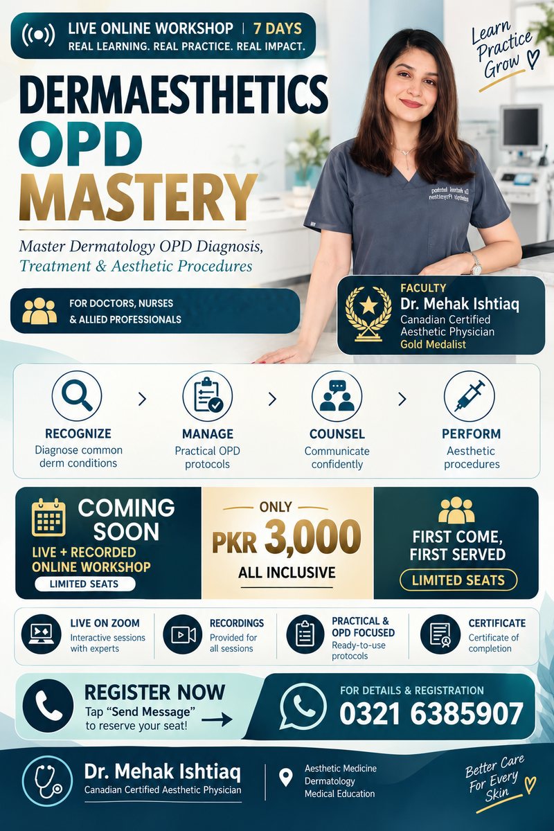
Try before you register

Sample lecture & notes

Sample lecture
Sample notes

Day 2 notes: Pigmentary disorders (preview). A preview of the notes you'll receive with this course.

Download sample notes (PDF)
FAQs

Common questions

When will this course start?

Dates will be announced soon. Register your interest and we will message you on WhatsApp first.

Who can join?

Doctors, nurses and allied health professionals. Procedures are discussed within each participant's legal scope of practice.

How is the course run?

Seven live classes on Zoom, 8–10 PM Pakistan time, usually on alternate days (not consecutive). Batches are kept small for individual attention, and every session is recorded.

What is the fee?

The fee is a fixed PKR 3,000 for the 7-session course. There is no early-bird offer.

Will I get a certificate?

Yes, a certificate of completion, issued within 7 days of the final class. This is clinical skills training, not a PMDC/HEC-recognised degree or postgraduate qualification.

Sample certificate of completion from Ashfaq Academy of Clinical Medicine, marked SAMPLE SAMPLE
Sample certificate of completion (tap to enlarge). Yours carries your name, the course dates, a certificate number, Dr Waqas Khan's signature and the institute seal. PDF
Will I get the recordings?

Yes. Every enrolled student gets 6 months of access to the session recordings, so a missed class is never a missed topic.

Is there a case-discussion group?

Yes. All members can join our free WhatsApp case-discussion group.

Do returning students get a discount?

Yes. If you have attended any of our courses or workshops before, you get a discount on your next one. Please ask us on WhatsApp for the current discount.

How do I get the Zoom link and recordings?

Each batch has its own private WhatsApp group. Zoom join links, recordings and case discussions are shared there. You are added once your payment is confirmed.

I live outside Pakistan. Can I join?

Yes, overseas students are welcome. 8 PM Pakistan time is 7 PM in the UAE, 6 PM in Saudi Arabia and 3 PM in the UK in winter (GMT), or 4 PM during British Summer Time. Register with your own country code and pay by JazzCash, Easypaisa or UBL bank transfer.

How do I register and pay?
  1. Fill in the registration form and choose your course. You will get a reference number such as AACM-261019-AB12.
  2. Send the course fee to one of our accounts: JazzCash: 03216385907 (Waqas Khan); Easypaisa: 03216385907 (Waqas Khan); UBL (United Bank Limited): account title Waqas Khan, account no. 0381318599799, IBAN PK33UNIL0109000318599799. Please write your reference number in the transfer note.
  3. Upload your payment receipt (image or PDF, up to 8 MB) with the transaction ID.
  4. We verify your payment and confirm your seat on WhatsApp. You can download a PDF of your registration straight away.

Courses marked 'Coming soon' only need a short 'register interest' form; there is no payment until dates are announced.

Course poster

Share this course

Tap the poster to view it full size, download it, or send it to a colleague on WhatsApp.

Dermaesthetics OPD Mastery poster: 7-day live online workshop, coming soon, PKR 3,000, with Mehak Ishtiaq, Canadian-certified aesthetic physician
Download poster